Back Of Skull Anatomy / Crown Of Head Conditions Injuries And More. Learn about the anatomy of the skull bones and sutures as seen on ct images of the brain. Human anatomy for muscle, reproductive, and skeleton. Synarthrodial joints, which allow no movement. Looking at it from the inside it can be subdivided into. Ct anatomy of skull, axial reconstruction, bone window.
Looking at it from the inside it can be subdivided into. Skull, skeletal framework of the head of vertebrates, composed of bones or cartilage, which form a unit that protects the brain and some sense organs. These joints fuse together in adulthood. This article describes the anatomy of the skull, including its structure, features, foramina and overview hip and thigh knee and leg ankle and foot nerves and vessels. Related posts of bone of back of skull.
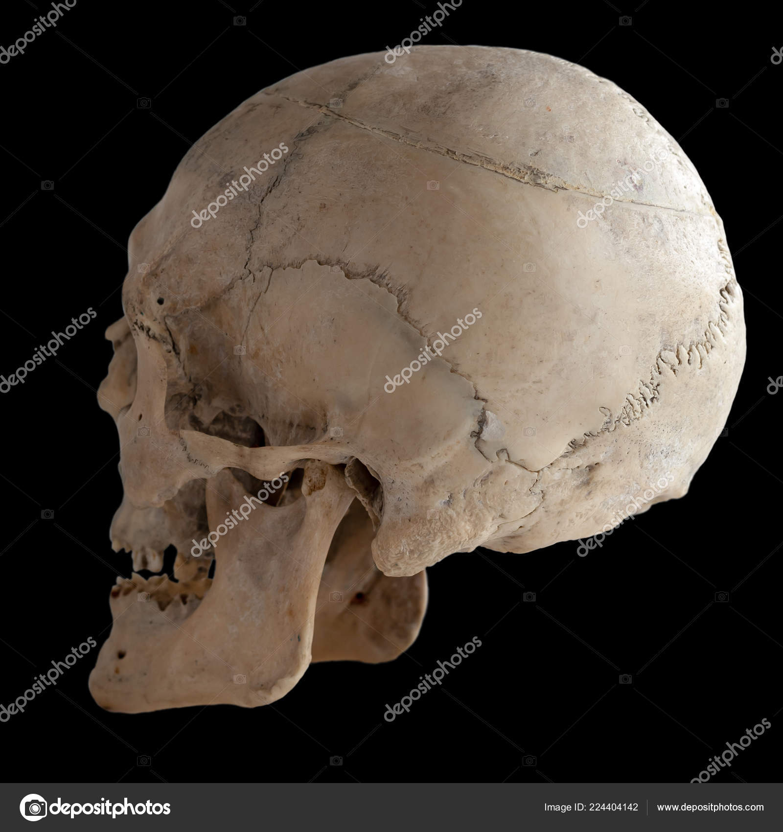
Anatomical Real Human Skull Closeup Angle Back Isolated Black Background Stock Photo Image By C Cobalt 70 224404142 from st4.depositphotos.com The skull also includes cartilage (put your finger on the tip of your nose and wiggle it) and ligaments (open and close your mouth if you want to use them). The skull or known as the cranium in the medical world is a bone structure of the head. The skull performs vital functions. Some bones give shape to the face, others protect the brain. Human skull from the front. Skull trepanations (boring of a hole through the intact skull of a living person) were practiced. Ct anatomy of skull, axial reconstruction, bone window. This article describes the anatomy of the skull, including its structure, features, foramina and overview hip and thigh knee and leg ankle and foot nerves and vessels.
Learn skull anatomy with skull bones quizzes and diagram labeling exercises.
Learn about the anatomy of the skull bones and sutures as seen on ct images of the brain. It is believed that trepanation was used to either relieve painful headaches, or to release demons from the skull. But it's not all bones! The skull begins to form prior to week 12 of embryogenesis. It supports and protects the face and the brain. It was then cleaned, adapted and polypainted this model is part of a comparison with the skull of a human. It is comprised of many bones, formed by intramembranous ossification, which are joined together by sutures (fibrous joints). In order to be light, the skull is made up by flat and irregular bones, and has hollow spaces called the sinuses. Overview, anterior skull base, middle skull base march 18, 2017. The cranium and mandible was exported from ct data. Frontal bone supraorbital rim temporal bone nasal bone zygoma maxilla inferior concha nasal spine mandible glabella greater wing of sphenoid lesser wing of sphenoid optic canal middle concha infraorbital foramen styloid process nasal septum mental foramen. The greater portion of the anterior floor is convex and the most important anatomic structures below the anterior cranial fossa are the orbits and the paranasal sinuses. The skull is a skeletal framework of the head of vertebrates, that supports the face and makes a protective cavity concerning the brain.
Skull contains both junction types: A thorough description is beyond the. The major sutures are the coronal suture, sagittal suture, lambdoid suture and squamosal sutures. Looking at it from the inside it can be subdivided into. The skull also incorporates the upper parts of the digestive (mouth) and respiratory tracts (nose).
Occipital Artery Anatomy Function And Significance from www.verywellhealth.com In order to be light, the skull is made up by flat and irregular bones, and has hollow spaces called the sinuses. It is believed that trepanation was used to either relieve painful headaches, or to release demons from the skull. Related posts of bone of back of skull. A thorough description is beyond the. Synarthrodial joints, which allow no movement. It is comprised of many bones, formed by intramembranous ossification, which are joined together by sutures (fibrous joints). Cranial cavity , cranial sutures. The skull base is the inferior portion of the neurocranium.
The skull also includes cartilage (put your finger on the tip of your nose and wiggle it) and ligaments (open and close your mouth if you want to use them).
Anatomy and physiology7.2 the skull. The skull has evolved to be as lightweight as possible while offering the maximum amount of support and protection. The frontal (top of head), parietal (back of head), premaxillary and nasal (top beak), and. So, the human skull consists of 23 bones. A thorough description is beyond the. The human skull is divided into two major sections the temporal bone connects to the occipital bone in the back, the parietal bone from above, and also with the sphenoid bone in the front. The skull base is the inferior portion of the neurocranium. This anatomic region is complex and poses surgical challenges for otolaryngologists and neurosurgeons alike. Some bones give shape to the face, others protect the brain. The bbc is not responsible for the content of external websites. Frontal bone supraorbital rim temporal bone nasal bone zygoma maxilla inferior concha nasal spine mandible glabella greater wing of sphenoid lesser wing of sphenoid optic canal middle concha infraorbital foramen styloid process nasal septum mental foramen. Human anatomy for muscle, reproductive, and skeleton. Home » drawing tutorials » basic drawing tutorials » skull anatomy.
Anatomy ▶ head and neck ▶ bones and cartilages ▶ skull. Bone of back of skull. Some bones give shape to the face, others protect the brain. They don't move and united into a single unit. Overview, anterior skull base, middle skull base march 18, 2017.
The Human Skull Anatomical Chart 9781587791673 Medicine Health Science Books Amazon Com from images-na.ssl-images-amazon.com They don't move and united into a single unit. Skull contains both junction types: It is believed that trepanation was used to either relieve painful headaches, or to release demons from the skull. Atlas of human skeletal anatomy. Anatomy and physiology7.2 the skull. Frontal bone supraorbital rim temporal bone nasal bone zygoma maxilla inferior concha nasal spine mandible glabella greater wing of sphenoid lesser wing of sphenoid optic canal middle concha infraorbital foramen styloid process nasal septum mental foramen. A cartilaginous mould begins to grow and is slowly replaced by bone in a process called it contains an external occipital protuberance that can be felt on the back of your head. The cranium and mandible was exported from ct data.
Anatomy and physiology7.2 the skull.
The skull or known as the cranium in the medical world is a bone structure of the head. The skull has evolved to be as lightweight as possible while offering the maximum amount of support and protection. Skull bones aren't fused together at birth. So, the human skull consists of 23 bones. It was then cleaned, adapted and polypainted this model is part of a comparison with the skull of a human. Related posts of bone of back of skull. They don't move and united into a single unit. The occipital muscle is cupped like a saucer to accommodate the back part of the brain. Overview, anterior skull base, middle skull base march 18, 2017. The major sutures are the coronal suture, sagittal suture, lambdoid suture and squamosal sutures. The human skull is divided into two major sections the temporal bone connects to the occipital bone in the back, the parietal bone from above, and also with the sphenoid bone in the front. The skull includes the upper jaw and the cranium. Ct anatomy of skull, axial reconstruction, bone window.
Share :
Post a Comment
for "Back Of Skull Anatomy / Crown Of Head Conditions Injuries And More"
:max_bytes(150000):strip_icc()/human-skull-with-veins-and-arteries--rear-view--1174640349-490cb7f8593945c4b1690b152e6a4074.jpg)


Post a Comment for "Back Of Skull Anatomy / Crown Of Head Conditions Injuries And More"